
Compatible with Evident (Olympus) epi-fluorescence OEM products
Translation: 3 — 5 axis stages allow scanhead rotation and translation in


TURN-KEY, FLEXIBLE, MULTIMODAL, COMPACT
The MPC-series combines the integrated design of our MPX-series with the flexibility of fluorescence microscopes.
It utilizes the Evident® (Olympus) fluorescence illuminator, which allows the users to add various parts from the Evident® (Olympus) universe, e.g. tube lens or filter wheels.
- Highly customizable
- 3-Photon and THG-ready
- Fully custom-designed laser scanning optics, achromatically compensated from 680nm to 1700nm, covering the full range of Ti:Sapphire lasers, fiber lasers as well as OPO/OPA/OPCPA systems up to 1700nm.
- Up to 4 ultra sensitive GaAsP PMTs with low dark count rate in epi or transmisson
- Compatible with Evident (Olympus) epi-fluorescence OEM products
- Open frame for later upgrades
- Optimized fluorescence detection through large collection angle and 2“ optics
Specifications
The MPC scan optics is fully custom designed and covers the entire range from 680nm to 1750nm. It is therefore well suited for 2P, 3P and 4P, as well as label free SHG, THG, CARS, SRS and FLIM imaging.
The MPC is compatible to all Prospective Instruments FSX ultrafast lasers, as well as suitable laser models from all other major ultrafast laser manufacturers.
(I) Multiphoton fluorescence imaging
Motorized laser power control
0.5 % - 100 %
Scan path
Galvo-galvo or resonant-galvo-galvo scanner*
Scan speed (galvo-galvo / resonant-galvo-galvo)
4.6 fps at 512 x 512 pixels
0.3 fps at 2048 x 2048 pixels
Pixel dwell time: 0.8 to 32 µs
0.3 fps at 2048 x 2048 pixels
Pixel dwell time: 0.8 to 32 µs
30 fps at 512 x 512 pixels
(8 kHz resonant galvo CRS8K)
Pixel dwell time: 44 ns
(8 kHz resonant galvo CRS8K)
Pixel dwell time: 44 ns
Field of view (FOV)
20 mm diagonal square (max) at the intermediate image plane
Beam diameter @ objective back aperture plane
20 mm
Point spread function
Depending on installed objective
Scan zoom (digitally via ScanImage)
1x to 99x
Scan size
Up to 2048 x 2048 pixels (Both bi- and unidirectional)
Multiphoton signal detection
Detection
Non-descanned fluorescence excitation detection, up to 4 PMTs, HDDs, SiPMs or SPADs in epi or transmission.
Collection optics
Extra-large highly efficient 2“ achromatically compensated detection optics, collection of fluorescence photons within +/- 12° from objective back arperture.
(II) Modality: widefield fluorescence imaging
Lightsource
White-light LED or up to 9 channel solid-state based illumination.
Filter set
Excitation, dichroic and emission filter sets individually selectable, matching illumination light source option.
Fluorescence camera
sCMOS monochrome, 6.5 µm pixel size, readout noise 2.1 med e-, q.e. up to 80 %, spectral range 370 nm - 1100 nm, dark current (typ.) 15 e- / pixel / s. All other major fluorescence camera models can be integrated.
Widefield options
Olympus OEM catalogue, such as binoculars, trinoculars, Köhler illumination, multiple camera ports. Wide selection of evident. Olympus of OEM catalogue possible.
Software
ScanImage Basic or Premium 2023 or higher
Laser scanning
ChromogazerTM
System monitoring & modality change
MS WindowsTM 10 64-bitProfessional
PC operating system
ImageJ (Fiji)
Image post-processing
Matlab
Scanimage API and aquisition scripts for fully autonomous imaging
Controller
Umbilical
Non-detachable umbilical between controller and scanhead, >2.0 m length
Embodyment
Stand-alone controller on wheels.
Cooling
No chiller, fully aircooled
Power
Single phase, 85 - 240 VAC, 10 A max (max 800 W total power consumption).
Built-in PC hardware
ATX gaming board, AMD Ryzen, 64 GB RAM, 500 GB SSD, 4 TB HDD, Quadro RTX GPU. High performance PC platform or better.
Size scanhead
50 cm x 40 cm x 15 cm (WxHxD)
Operating enviroment
18°C - 27°C. Extended operating conditions available.
Storage temperature
-15°C to +50°C.
Humidity
10% - 90% (non condensing)
Objectives
Turret
3-positions, motorized & software controlled
Turret threadings
M32 x 0.75
Objectives
Scan optics compatible to all major objective manufacturers. Optimized for state-of-the-art high-NA objectives.

Download full specification
Broad range of applications
- 2D/3D/4D imaging
- In-vitro & in-vivo
- Whole slide imaging
- Label-free & IHC/ICC
- Non-destructive
- Deep tissue imaging
-
Pathology & Cancer
-
Neuroscience
-
Optogenetics
-
Tissue Engineering & Bioprinting
-
Spheroids/Organoids
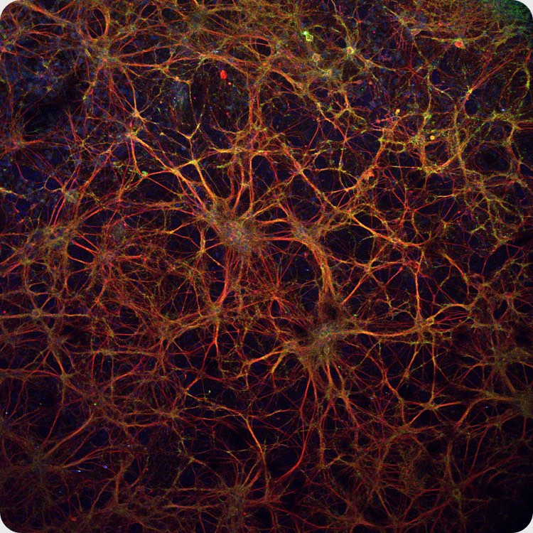
Gallery


SOFTWARE INTEGRATIONS
Our software allows for the collection of more reliable data in less time. Our Windows 10 scripts are customized to automate workflows and integrate external image analysis programmes.


Get a quote
We’ll be more than glad to tell you all about our products.
Just fill out the form and we’ll get in touch with you shortly.
Prospective Instruments LK OG Bildgasse 18,6850 Dornbirn,
Austria

Partners of myclimate.org and
Climate Neutrality Alliance 2025.